Lowe, Jesse
(2017)
SIS-ECM Scaffold Remodels Into a TMJ Disc Analogue.
Doctoral Dissertation, University of Pittsburgh.
(Unpublished)
Abstract
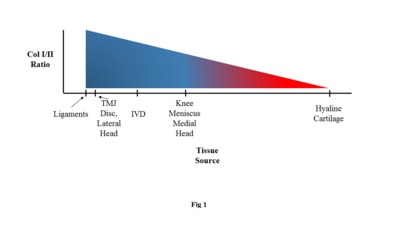
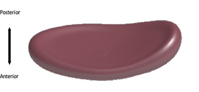
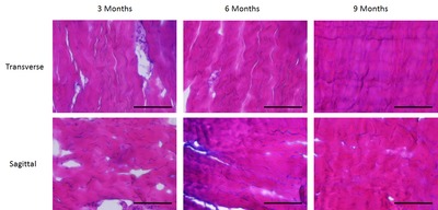
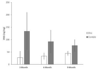
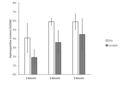
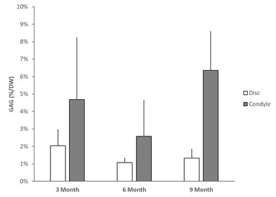
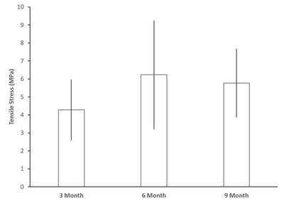
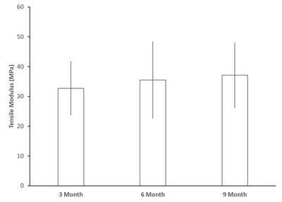
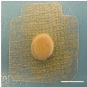
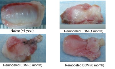
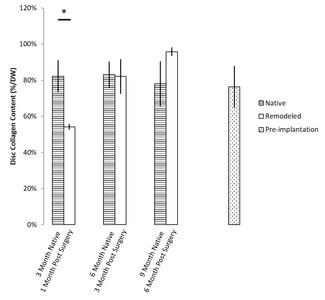
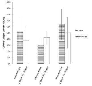
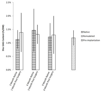
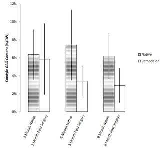
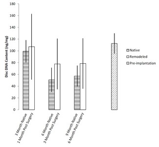
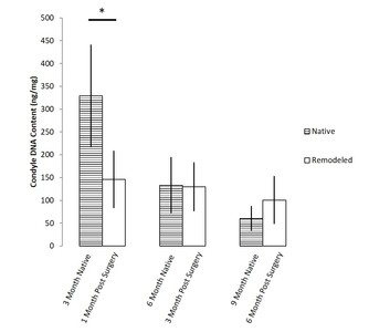
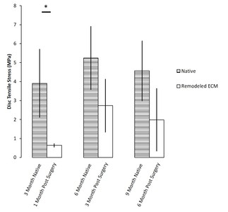
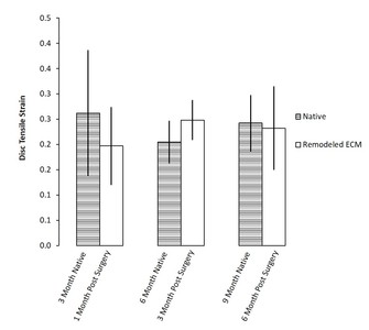
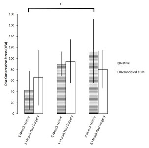
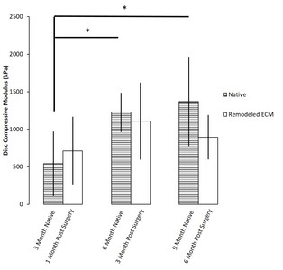
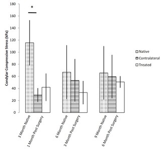
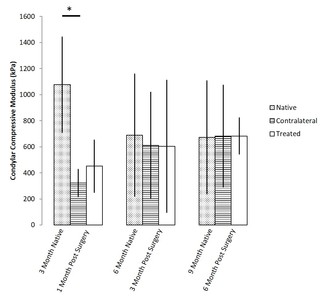
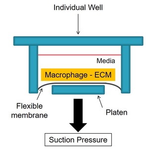
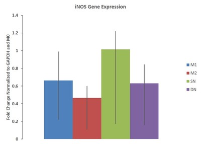
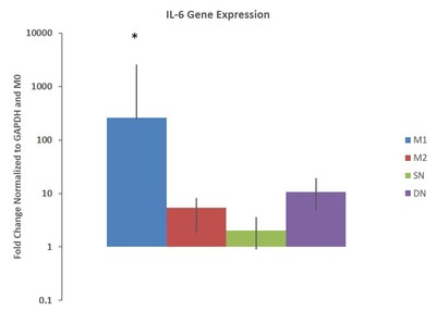
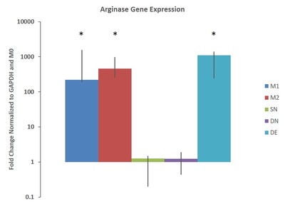
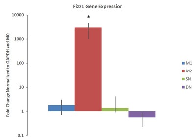
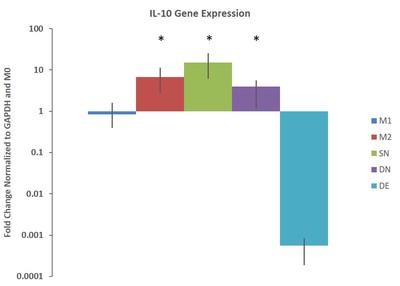
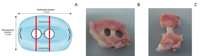
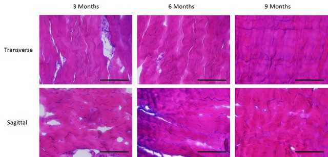
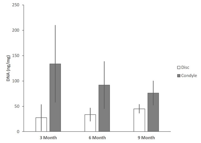
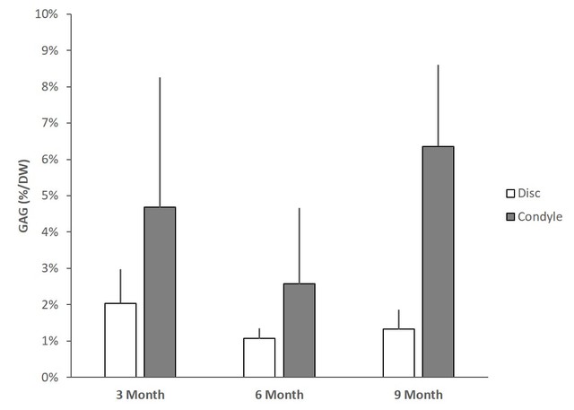
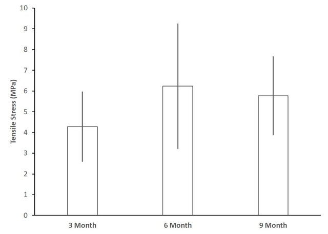
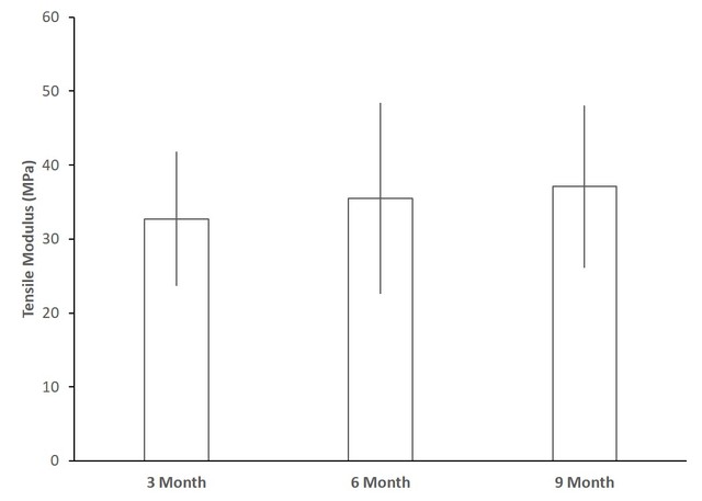
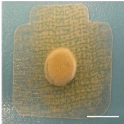
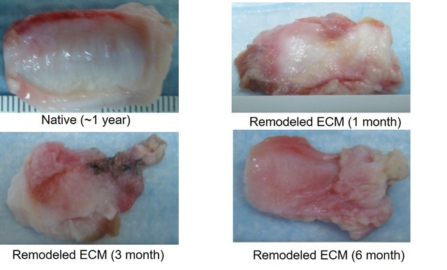
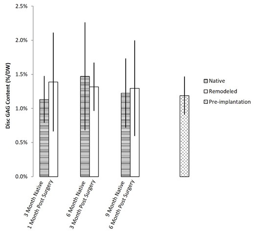
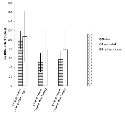
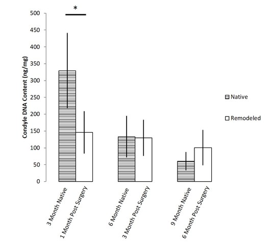
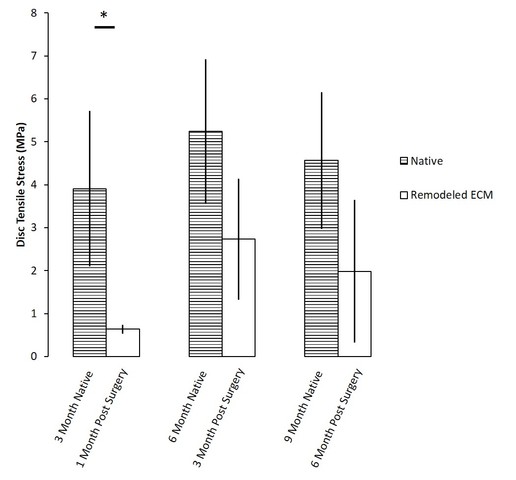
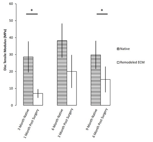
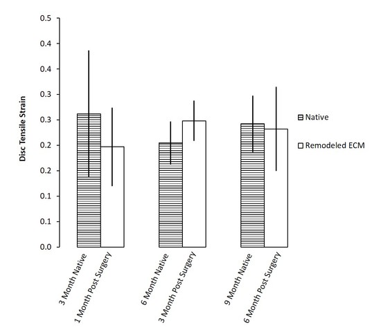
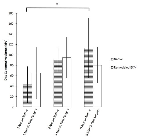
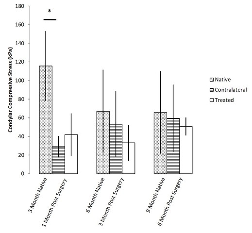
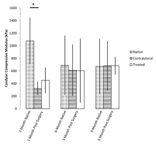
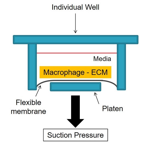
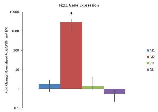
The temporomandibular joint (TMJ) disc is a fibrocartilaginous tissue located between the condyle of the mandible and glenoid fossa and articular eminence of the temporal bone, forming the TMJ. Damage or derangement of the TMJ disc can require surgical removal, but current autograft replacements will generally resorb within a year, highlighting the need for long term solutions. Extracellular matrix (ECM) scaffolds have shown potential as regenerative medicine graft replacements. Toward this end, the work described in this thesis provides a systematic in vivo approach for the use of small intestine submucosa (SIS) ECM in replacing the porcine disc. Initial studies focused on the effect of growth on the biochemical and biomechanical properties of the porcine TMJ disc and condylar cartilage by characterizing these properties in native pigs at 3, 6, and 9 months. It was determined that growth had no significant effect on the properties of the TMJ disc and condyle. Once baseline native properties were determined, SIS-ECM scaffolds were implanted unilaterally in a porcine TMJ following disc removal and allowed to remodel for 1, 3, and 6 months post implantation. The excised remodeled scaffolds and associated condyles were then characterized and compared against age matched control TMJ discs and condyles. It was determined that the remodeled scaffolds were able to recapitulate native biochemical properties and achieve 50% of the native tensile properties within 3 months post-implantation. The effect of implantation of the ECM scaffolds on the properties of the condylar cartilage seemed to be the same as no implantation, but both healed. Finally, an in vitro investigation into the effect of ECM and mechanical stimulation on macrophage modulation was performed to provide insight on early ECM scaffold remodeling. It was determined that mechanical stimulation (4 hours of 5% strain at 1 Hz) did not have a significant effect on MO macrophage phenotype towards M1 or M2, while the combination of ECM and mechanical stimulation caused macrophages to display a M2 phenotype. The success of these studies suggest the efficacy of the SIS-ECM scaffold as a potential tissue engineered graft replacement for the damaged TMJ disc.
Share
| Citation/Export: |
|
| Social Networking: |
|
Details
| Item Type: |
University of Pittsburgh ETD
|
| Status: |
Unpublished |
| Creators/Authors: |
|
| ETD Committee: |
|
| Date: |
30 June 2017 |
| Defense Date: |
3 April 2017 |
| Approval Date: |
26 September 2017 |
| Submission Date: |
30 June 2017 |
| Access Restriction: |
3 year -- Restrict access to University of Pittsburgh for a period of 3 years. |
| Number of Pages: |
147 |
| Institution: |
University of Pittsburgh |
| Schools and Programs: |
Swanson School of Engineering > Bioengineering |
| Degree: |
PhD - Doctor of Philosophy |
| Thesis Type: |
Doctoral Dissertation |
| Refereed: |
Yes |
| Uncontrolled Keywords: |
Tissue Engineering, Biomechanics, Extracellular Matrix, Temporomandibular Joint |
| Date Deposited: |
26 Sep 2017 16:56 |
| Last Modified: |
26 Sep 2020 05:15 |
| URI: |
http://d-scholarship.pitt.edu/id/eprint/32664 |
Metrics
Monthly Views for the past 3 years
Plum Analytics
Actions (login required)
 |
View Item |


![[img]](http://d-scholarship.pitt.edu/32664/33/image1.png)
![[img]](http://d-scholarship.pitt.edu/32664/18/image2.png)
![[img]](http://d-scholarship.pitt.edu/32664/14/image3.png)
![[img]](http://d-scholarship.pitt.edu/32664/23/image4.png)
![[img]](http://d-scholarship.pitt.edu/32664/24/image5.jpeg)

![[img]](http://d-scholarship.pitt.edu/32664/30/image6.png)
![[img]](http://d-scholarship.pitt.edu/32664/17/image7.jpeg)
![[img]](http://d-scholarship.pitt.edu/32664/6/image8.jpeg)
![[img]](http://d-scholarship.pitt.edu/32664/8/image9.jpeg)
![[img]](http://d-scholarship.pitt.edu/32664/26/image10.jpeg)
![[img]](http://d-scholarship.pitt.edu/32664/9/image11.jpeg)
![[img]](http://d-scholarship.pitt.edu/32664/19/image12.jpeg)
![[img]](http://d-scholarship.pitt.edu/32664/13/image13.jpeg)
![[img]](http://d-scholarship.pitt.edu/32664/16/image14.jpeg)
![[img]](http://d-scholarship.pitt.edu/32664/4/image15.jpeg)
![[img]](http://d-scholarship.pitt.edu/32664/31/image16.jpeg)
![[img]](http://d-scholarship.pitt.edu/32664/22/image17.jpeg)
![[img]](http://d-scholarship.pitt.edu/32664/3/image18.jpeg)
![[img]](http://d-scholarship.pitt.edu/32664/20/image19.jpeg)
![[img]](http://d-scholarship.pitt.edu/32664/5/image20.jpeg)
![[img]](http://d-scholarship.pitt.edu/32664/34/image21.jpeg)

![[img]](http://d-scholarship.pitt.edu/32664/28/image22.jpeg)
![[img]](http://d-scholarship.pitt.edu/32664/15/image23.jpeg)
![[img]](http://d-scholarship.pitt.edu/32664/10/image24.jpeg)
![[img]](http://d-scholarship.pitt.edu/32664/25/image25.jpeg)
![[img]](http://d-scholarship.pitt.edu/32664/2/image26.jpeg)
![[img]](http://d-scholarship.pitt.edu/32664/12/image27.jpeg)
![[img]](http://d-scholarship.pitt.edu/32664/29/image28.jpeg)
![[img]](http://d-scholarship.pitt.edu/32664/7/image29.jpeg)

![[img]](http://d-scholarship.pitt.edu/32664/11/image30.jpeg)
![[img]](http://d-scholarship.pitt.edu/32664/32/image31.jpeg)
![[img]](http://d-scholarship.pitt.edu/32664/21/image32.jpeg)
![[img]](http://d-scholarship.pitt.edu/32664/27/image33.jpeg)


